All of us have heard of tonsil surgery once in our lifetime. But do any of us have any idea how it is done? I myself have absolutely no idea until four years of medical school.
The surgery is done for those with frequent episodes of tonsil infection, enlarged tonsils causing snoring & airway compromise, suspected cancer cases and few other rare conditions.
Surgery is done under general anaesthesia, meaning they will be fully asleep & pain free during the procedure. The patient, a boy as depicted here, is given a mask to breathe till he is fully asleep (image 1).
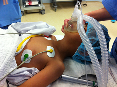
Image 1: going under general anaesthesia
A tube is then inserted into the mouth (image 2) into the airways to provide oxygenation (endotracheal tube) and connected to a machine for him to breathe (ventilator). The surgery will be done with the mouth open with the surgeon sitting facing the top of the head.
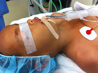
Image 2: tube inserted inserted through the mouth, same area as surgical field
There are plenty of methods for tonsillectomy. In my hospital, we use sharp instruments (image 3) combined with diathermy (heat method of dissection). Other centres may use different instruments.
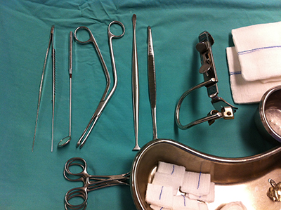
Image 3: part of the instruments used for tonsillectomy
The tonsil is approached from the open mouth. Therefore, no skin incision is made and no scars will be present externally (image 4)
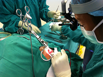
Image 4: Surgeon operates from the top aided with the headlight for a brighter view of the hollow cavity
Since it is seen from the top, the tonsil appears upside down as seen in this image 5. A small amount of adenoid tissue is taken sometimes in children, approached from below the soft palate. Adenoid is similar to the tonsil, but present at the back area of the nose (nasopharynx)
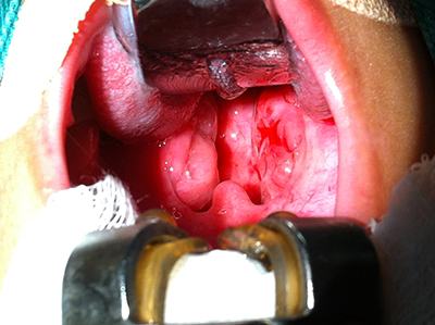
Image 5: enlarged tonsils noted parted by the mouth retractor pressing on the tongue above. Adenoid is not seen, hidden below the soft palate.
After removal, the bleeding areas are secured with diathermy (image 6 & 7). This is essential as he may bleed in the ward if he coughs or cries loudly. The entire procedure takes roughly 30 minutes, depending on case difficulty and the surgeon’s skills & experience.
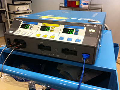
Image 6: the diathermy machine used in surgery for dissection and securing bleeding points.
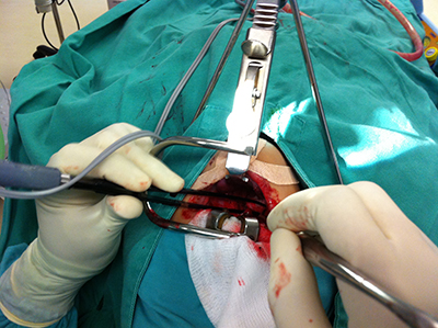
Image 7: the diathermy attachment on the left hand and suction on the right hand to clear the blood.
The tissue removed is then set for lab examination for confirmation of diagnosis (image 8). The patient usually stays in the ward for another day after surgery. He may go home if there is no bleeding, no fever and able to take adequate amounts of soft diet orally.
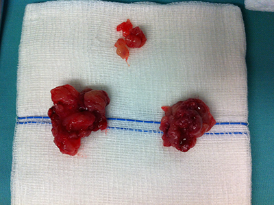
Image 8: the 2 large specimens below shows left & right tonsils while the smaller one above is the adenoid
The patient will be given antibiotics, painkiller & mouth gargle. We usually will review patients weeks later to review specimen result and assess possible complications such as residual symptoms, voice change, swallowing difficulties and altered taste.
Dr Ahmad Nordin is an Ear Nose and Throat surgeon currently working in Sabah. Find out more about him on The Team page.
References:
- Synopsis of operative ENT Surgery, Bingham & Hawthorne
- Diseases of the Ear, Nose and Throat 4th Edition PL Dhingra
[This article belongs to The Malaysian Medical Gazette. Any republication (online or offline) without written permission from The Malaysian Medical Gazette is prohibited.]